Research Topics of Rüdiger Berger

Currently, my research is focussing on two topics: (a) Understanding and controlling of lateral adhesion forces of liquid drops that slide over surfaces. (b) Understanding charge extraction in solar cells and solid-state battery materials on a nanometer scale using electrical modes of scanning force microscopy.

Friction force measurements of drops

We built up a method that allows the measurement of forces required to slide sessile drops over surfaces. The forces were measured by means of a deflectable glass capillary stuck in the drop (video close by). Deflections of this capillary are detected by imaging with a CCD camera. The drop's friction force instrument (DoFFI - previously called DAFI) allowed the investigation of the static and dynamic lateral friction force of water drops. The movement of the drop relative to the surfaces enabled us to resolve the pinning of the three-phase contact line to individual defects [1]. This pinning force is identical to the force calculated by the deformation of the three phase contact line base on a model reported by Joanny and deGennes [2].

The DoFFI is a suitable tool for characterizing friction forces on hydrophobic surfaces. In particular, a 2-dimensional scanning of the samples allows us to resolve different wetting phenomena of surfaces spatially. Thus, DoFFI is a novel scanning probe method, named scanning drop friction force instrument (sDoFFI) [3]. In sDoFFI the drop acts as the probe. 
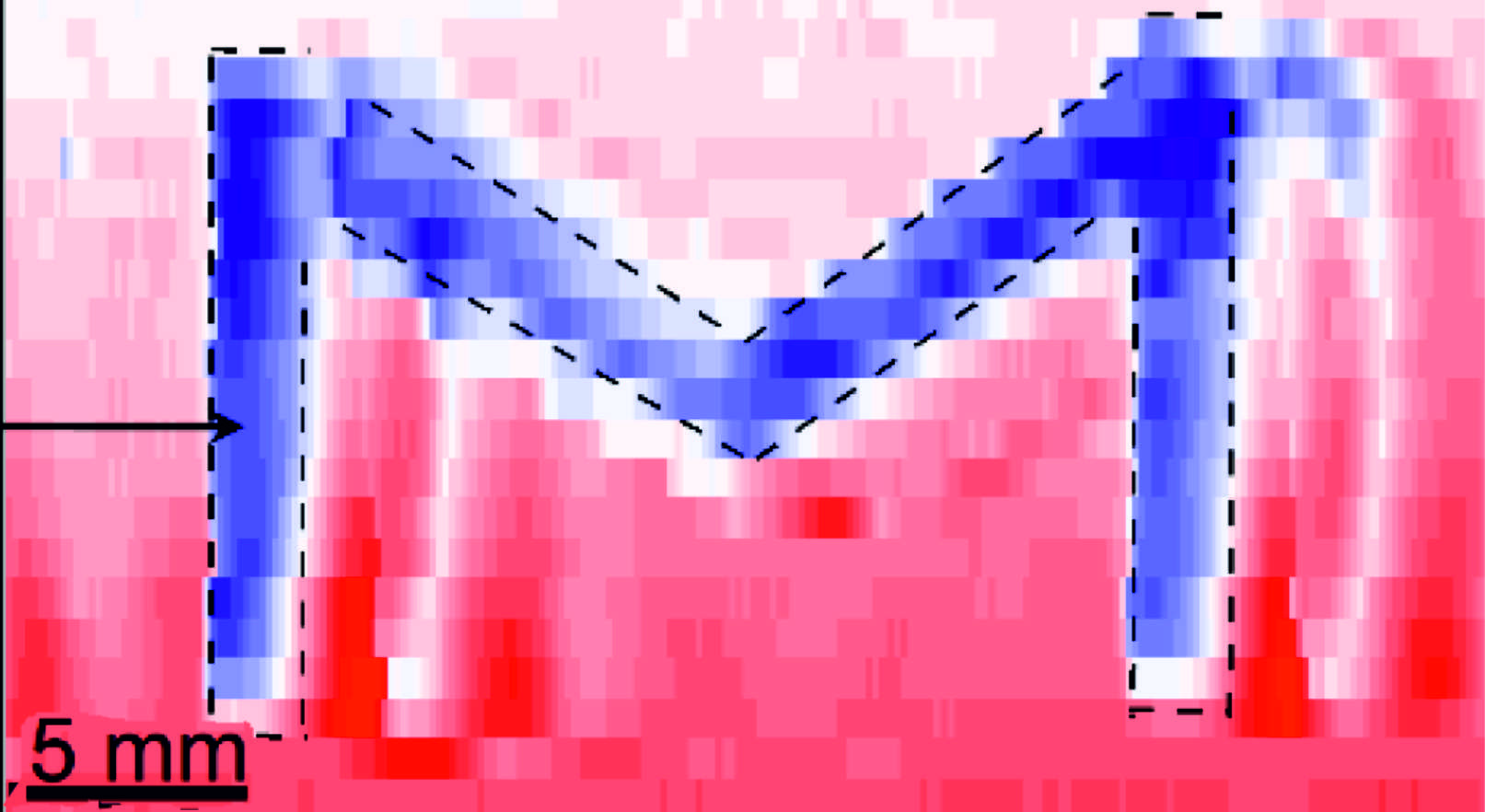
sDoFFI is applicable for quality control of surfaces made by large-scale industrial processes. The close sDoFFI map shows the friction force resultig from a M-feature made by CVD of OTS and PFOTS molecules. For more details visit the DoFFI homepage.

We used DoFFI to investigate the "static" and a "kinetic" regime of sliding drops [4] and to characterize charging of sliding drops and study the interaction between transport and wetting processes within the collaborative research center 1194. Recently, we investigated how contamintations are removed from surfaces by sliding drops. We monitored the removal of individual contaminant particles on the micron scale by confocal microscopy while drops are kept in position by DoFFI. We correlate the space- and time-resolved information with measurements of the lateral friction force of the sliding drop [5]. In a recent study we show that DoFFI is a better alternative to characterize material properties compared to measurements of the onset of motion, e.g. by tilted plane. The reason is that the static friction force can be tuned by over 30% by pre-shaping the drop. In contrast to static friction, kinetic friction is independent of pre-shaping the drop, i.e. the drop history [6].

References:
[1] Dynamic Measurement of the Force Required to Move a Liquid Drop on a Solid Surface, D. W. Pilat et al., Langmuir (2012).
[2] Pinning forces of sliding drops at defects, A. Saal et al., Europhys. Lett. (2022).
[3] Scanning Drop Friction Force Microscopy, Chirag Hinduja et al., Langmuir (2022).
[4] How drops start sliding over solid surfaces, Nan Gao et al., Nature Physics (2018).
[5] When and how self-cleaning of superhydrophobic surfaces works, F. Geyer et al., Science Advances (2020).
[6] Tuning drop friction, Alexandre Laroche et al., Droplet (2023).

Local electrical current transport through interfaces

Interfaces play a major role in electrical devices. In case interfaces are heterogenous the electrical current that can flow through the interface locally varies. Such variations can be probed by conductive mode (cSFM). In cSFM the surface is typically scanned in contact mode. In addition an electrical potential (Us) is applied and the local current between tip and sample surface (Itip) is measured. However the operation of contact mode SFM is often destructive for soft matter surfaces. Therefore, we have developped and applied non-destructive modes based on torsion force microscopy [1],[2] and force distance based modes [3] (peak force tapping or quantitative imaging).
These modes were used to quantitify the current flow along individual nano-pillars made from a thermally cross-linked triphenylamine-derivate semiconductor [1] and made from P3HT and PCBM [3] (see also image). Furthermore, we studied TiO2 anatase thin films which were UV-ozone treated [4]. We found that the latter is an efficient method to increase the conductance through the film by more than one order of magnitude. The increased conductance of TiO2 anatase thin films results in a 2 % increase of the average power conversion efficiency (PCE) of methylammonium lead iodide based perovskite solar cells. PCE values up to 19.5 % for mesoporous solar cells are realized. Using cSFM we probed the conductance of homo- and blended conjugated polymers in confined in nanostructures. The resulting structures lead to high charge mobility along vertical direction for both homo- and blended conjugated polymers. We found a more than two orders of magnitude enhanced charge mobility along vertical direction [5].

References:
[1] Mapping of Local Conductivity Variations on Fragile Nanopillar Arrays by Scanning Conductive Torsion Mode Microscopy, Stefan A.L. Weber et al., Nano Letters, 10, 1194 - 1197 (2010). Langmuir 28, 16812-16820 (2012).
[2] Electrical tip-sample contact in scanning conductive torsion mode, Stefan Weber, Rüdiger Berger, Applied Physics Letters 102, 163105 (2013).
[3] Controlled Mutual Diffusion between Fullerene and Conjugated Polymer Nanopillars in Ordered Heterojunction Solar Cells, Jongkuk Ko et al., Advanced Materials Interfaces, 1600264 (2016).
[4] Removal of Surface Oxygen Vacancies increases Conductance through TiO2 Thin Films for Perovskite Solar Cells, Alexander Klasen et al., Journal of Physical Chemistry C (2019).
[5] Control of Electronic Properties of Functional Organic and Inorganic Materials Through Nano-Confinement, Jongkuk Ko, Rüdiger Berger, Hyemin Lee, Hyunsik Yoon, Jinhan Cho,and Kookheon Char, Chem. Soc. Review 50, 3585-3628 (2021)

Microcantilever Sensors

... a research area, where I am not very active an more.


An essential part of scanning force microscopy is a micromechanical cantilever sensor (MCS) which transduces a force acting on the tip into a deflection. Forces of pico newtons can be measured which correspond to a sub-nanometer deflection of the MCS. However, not only forces acting on the tip lead to a deflection, also expansive or contractive forces acting on one side of the cantilever surface result in a bending. For example this is the case for a swelling or a phase change of thin polymer films, which have been deposited on one side of a MCS. In addition, tensile and compressive surface stress changes arise when molecules specifically adsorb on one side of the cantilever surface. A review of MCS operating modes and their applications can be found in an article published in Materials Today.

In particular, the surface stress changes can be measured in liquids which is a requirement for most biochemistry applications. In the field of biotechnology, DNA hybridisation between self complementary strands leads to conformational changes which result in a cantilever sensor bending. In addition, polymer materials are very attractive as responsive coatings for various sensing application [1], [2]. Beside sensing, the MCS technique can be applied for material characterization. In cooperaton with Prof. Dr. Akiko Itakura (NIMS) and Prof. Dr. Masaya Toda (Univ. of Tohoku), we analyzed the response of MCS that are coated with polymers in different solvents. Hereby the Young's module of the polymer can be calculated from the bending response [3].
The cantilever sensors are very small (typically 0.5 µm thick, 50 µm wide and 500 µm long). This offers the possibility to arrange several of these cantilever sensors in an array on a single chip. Hereby, experimental noise can be reduced by averaging signals or the response of an analyte (e.g. mixture of substances) to different coated sensors can be studied. The cantilever sensor technique is useful for studying tiny amounts of materials that are expensive to be produced in large quantities.
Currently, we develop methods to measure mass changes of samples that are attached to the cantilever's end. Two phenomena can be investigated: (a) changes in mass upon heating of the sample. Such a method is called (micromechancial) thermogravimetry and we aim to analyze biominerals within a DFG project in cooperation with Prof. Dr. Filipe Natalio from the Weizmann Institute [4]. The movie displays a heated zeolite crystal that is filled with a yellow dye. The dye excapes at 200 °C (i.e. at about 15 seconds in the movie) and leaves a transparent zeolite behind after cooling down. (b) changes in mass of polymers upon exposure of the samples to solvents. Here we would like to develop a fast and reliable method to determine the mechanical properties of soft matter materials [5].


References:
[1] Thin Polyelectrolyte Multilayers Made by Inkjet Printing and Their Characterization by Nanomechanical Cantilever Sensors, Masaya Toda et al., Journal of Physical Chemistry C 118, 8071 - 8078 (2014).
[2] Simplifying cantilever sensors: Segmental analysis, a way to multiply your output, Jannis W. Ochsmann et al., Sensors and Actuators B 177, 1142 - 1148 (2013).
[3] Effective Young's Modulus Measurement of Thin Film Using Micromechanical Cantilever Sensors, Akiko N. Itakura et al., Japanese Journal of Applied Physics, 52, 110111 (2013).
[4] Pico-thermogravimetric material properties analysis using a diamond cantilever beam, Ioana Voiculescu et al., Sensors and Actuators A, 271, 356-363 (2018).
[5] Young's modulus of plasma-polymerized allylamine films using micromechanical cantilever sensor and laser-based surface acoustic wave techniques, Masaya Toda et al., Plasma Process Polym., e1800083 (2018).

The projects are integrated in the research at the Max Planck Institute for Polymer Research in particular in the science perfromed in the department Physics of Interfaces of Prof. Dr. H.-J. Butt.